My patients ask me what approach I will be using when I replace their hip. I inform them I will be using the mini-posterior approach and not the anterior approach. I explain to them that both approaches produce excellent long-lasting results.
One of the most important factors is with the Posterior approach it can be extended, compared to the Anterior approach – extended meaning that the incision can be made longer to expose underlying tissues that can then be released to correct an underlying condition. An example, if during preparation of the femur a fracture occurs, the fracture can more easily be defined and fixed using a Posterior approach.
The Posterior approach represents a continuum or spectrum of approaches. From the Mini Posterior to Standard Posterior to Extended Posterior which is used when more femoral exposure is needed, such as when performing an Extended Trochanteric Osteotomy (ETO). The ETO is an important technique when opening up the upper femur, typically to remove a hip stem during revision surgery.
For many years I’ve used what I call a Mini Posterior approach which is basically the Standard Posterior approach but done through a smaller incision with significantly less underlying soft tissues release or dissection compared to the Standard Posterior approach. The exact definition of what constitutes a Mini Posterior approach is not fully agreed upon among surgeons or in medical literature. Some emphasize the length of the incision being <10 cm. I think the length of the incision is not nearly as important as which specific structures underneath the incision are divided or released.

What I now prefer is the SPAIRE technique :
S – Spairing
P – Piriformis
A – and
I – Internus (obturator)
R – Repairing
E – Externus (obturator)
Technically the SPAIRE technique is more difficult to perform but it is also the most muscle and tissue-sparing of the Mini Posterior Approaches and results in fewer tendons being damaged than any other approach, including the Direct Anterior Approach (DAA) or the Direct Superior Approach.

A person’s hip has a number of small muscles that insert above and in back of a hip joint, just on the outside of the hip joint capsule, collectively these are called “short external rotators.” Visualize these as a cloak of muscles that overlie the hip joint capsule. If we still “walked on all fours” they would be analogous to the rotator cuff muscles of our shoulder. Historically we relegated their function in our hip to being essentially vestigial (something no longer important) while in some animals assuming a more important function, for instance in the kangaroo. More recent studies are now recognizing that they do play an important role in helping stabilize our hips and assisting certain motions, particularly when the hip is flexed.
The Standard Posterior approach releases all of the “short external rotators” from the upper posterior femur as well as the gluteus maximus insertion into the femur. The Mini Posterior approach releases less and typically these structures are repaired back to bone with the hip capsule much more robustly than was done historically. The SPAIRE technique is a Mini Posterior approach and is what I do now. With this technique, only one tendon is released off the upper femur.
An interval between lowest external rotator muscles is developed and the cuff of muscles above are then elevated off the hip joint capsule. The hip joint capsule is opened under direct vision. Prior to doing this, the sciatic nerve is visualized so the risk of injury during the procedure is all but nullified because you can see it and monitor its safety throughout the procedure.
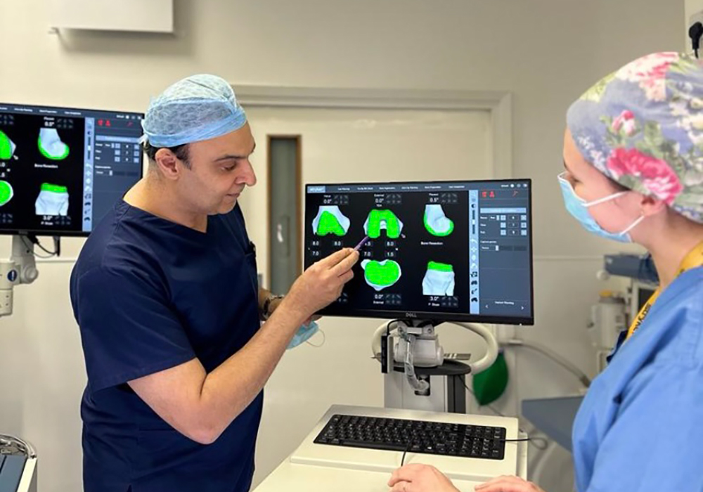
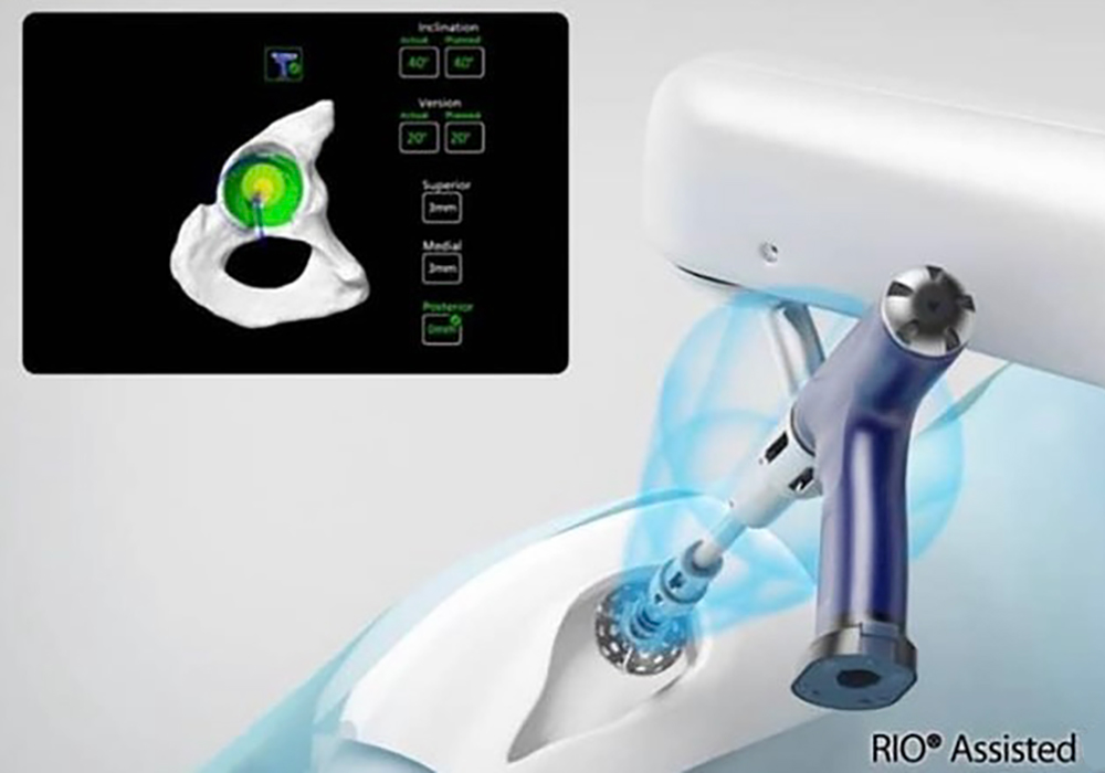
The SPAIRE technique allows for a robust anatomic capsular closure and repair of the inferior muscle cloak. The support provided by this natural cloak of overlying external rotator muscles and tendons as well as the hip joint capsule results in vastly improved stability against dislocation. This can be used with Mako Robotic Assisted Surgery which improves the accuracy of component postioning. This combination I was trained by Professor John Timperley in Exeter and we both believe this is the leading combination in hip surgery available.
Prior to my adopting the SPAIRE technique and after completing the hip reconstruction, I could clearly visualize the ball position in the new socket. Now I can see only the bottom third of the ball or less because the intact short external rotators and underlying hip joint capsule are covering the rest. When a hip dislocates posteriorly, that is it come out of the socket in back, the ball goes up and out (posteriosuperior). This posterior superior area surrounding the hip joint is now covered by the patient’s own hip capsule and intact short external rotator muscles and hence prevents the ball moving out in this direction.
Because this cloak of short external muscles is intact, their normal function is preserved, and the risk of dislocation is also less. Honestly, our risk of dislocation during a pre-SPAIRE Mini Posterior approach was also wonderfully low but now with this enhanced stability, I’ve discontinued all the classic post-operative hip precautions with my patients. Patients are encouraged to simply listen to their bodies and resume all of their usual activates when they feel comfortable.
My patients who had their first hip reconstructed by me with using my pre-SPAIRE Mini Posterior technique and now have had their second THR implanted using the SPAIRE technique are routinely asking what’s different? They tell me how they feel even better and are getting well even quicker. They get rid of their cane and any residual limp even sooner than before. This is anecdotal but for a clinician, powerful. I honestly don’t think long term results will be any different, but if someone can feel better quicker and not have restrictions, then I think it’s a positive move forward.
To book a consultation or for further information, contact:
Mr Nadim Aslam, Orthopaedic Robotic Surgeon, of the Worcestershire Knee & Hip Clinic
✉ [email protected]
☏ 01905 362003


